Veterinary Models
| Products | Item # | Information |
|
|
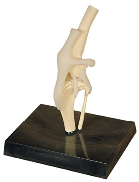
GPI1050 | Canine Knee
Average size dog knee with femur, fibula, patella and tibia bones, lateral
and medial meniscus, anterior and posterior cruciate ligaments, plus 6
more ligaments and tendons. 2" x 3-1/2" x 7"
PRICE: $81.80 |
|
|
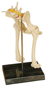
GPI1060 | Canine Pelvis (Hip) Model
Average size dog pelvis that features both normal and osteoarthritic bone, body of ilium, greater trochanter, head in acetabulum, herniated disk, neck of femur, nerve, sacrum, and third trochanter. 7" x 5-1/2" x 11"
PRICE: $110.00 |
|
|
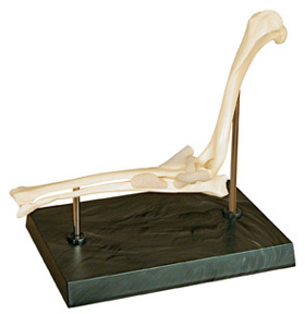
GPI1070 | Canine Elbow
Healthy left elbow of average size dog includes: humerus, radius and ulna bones; plus 6 ligaments. 9-1/2" x 1-3/4" x 7-1/2"
PRICE: $79.90 |
|
|
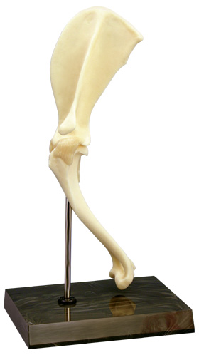
GPI1075 | Canine Shoulder
PRICE: $84.70 |
|
|
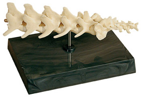
GPI1080 | Canine 5-piece Vertebrae
Vertebral column of average size dog features 5 lumbar vertebrae and discs, caudal (tail) vertebrae and sacrum. 8" x 2" x 2"
PRICE: $79.90 |
|
|
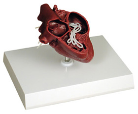
GPI1150 | Canine Heart
Cut-away view of an average size dog heart containing heartworm parasite. 3" x 3-1/2" x 4-1/4"
PRICE: $74.40 |
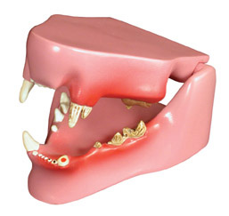 |
GPI1190 | Feline Jaw
Average size feline jaw depicts healthy teeth on the right side and diseased/damaged teeth on the left. The eight pathologies featured are: fractured canine, periodontal disease, tarter accumulation, plaque, gingivitis, worn incisors, retained deciduous tooth and missing premolar. The jaw may be opened, closed and separated for closer study.
PRICE: $79.90 |
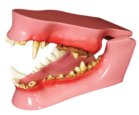 |
GPI1195 | Canine Jaw
Average size canine jaw depicts healthy teeth on the right side and diseased/damaged teeth on the left. The nine pathologies featured are: fractured canine, periodontal disease, tarter accumulation, plaque, gingivitis, worn incisors, retained deciduous tooth, gingival recession and missing premolar. The jaw may be opened, closed and separated for closer study.
PRICE: $85.50 |
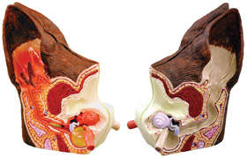 |
GPI1200 | Canine Ear
This 2-sided, average size, canine ear depicts: a normal side with cochlea, auditory tube, tympanic bulla, middle ear cavity, tympanic membrane, horizontal canal, vertical canal, auricular cartilage, pinna and temporalis muscle. The diseased side illustrates: inflamed inner ear structures, inflammatory exudate in tympanic bulla, ear canal with partial occlusion from cellular hyperplasia, inflammatory exudate and an inflamed outer ear. 4.75" x 2.75" x 2"
PRICE: $75.40 |
|
|
| Copyright © 1998-2009 a2zmed.com., All Rights Reserved TEL(804) 378-0077 FAX(804) 378-0997 |
| Use of this site indicates that you accept Terms and conditions, |
| Terms of Use and Privacy Policy of www.a2zmed.com |
| Send mail to sales@a2zmed.com with questions or comments about this web site. Last modified: 01/06/2009 |